
Services / Echocardiogram
Echocardiogram
A detailed ultrasound that shows the structure and function of your heart — valves, chambers, and blood flow — without radiation.
OHIP-insured with requisition
What it is
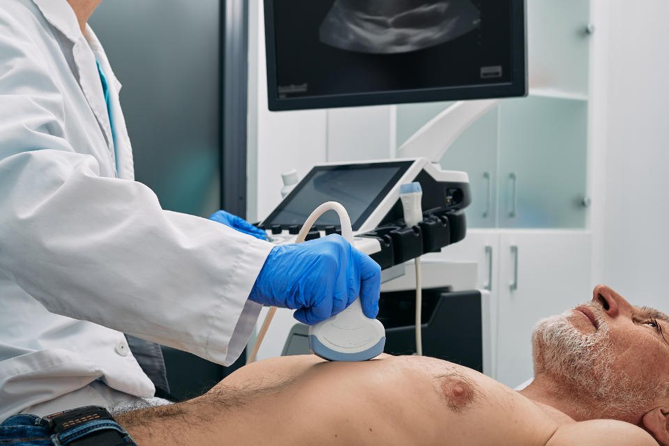
An echocardiogram (echo) is an ultrasound of the heart. A cardiac sonographer places a small probe on your chest with a dab of gel. Sound waves bounce off heart tissue and blood, and a computer turns those echoes into a live, moving picture. Colour and Doppler imaging let us measure how blood is flowing through your valves and chambers.
Why it is ordered
- Investigate chest pain, shortness of breath, swelling, or a murmur
- Assess valve disease, heart-muscle function, and chamber size
- Follow known heart conditions or check the heart before a procedure
- Screen when there is a strong family history of cardiomyopathy
Before your visit
- 01No fasting or special preparation is required
- 02Take your medications as prescribed unless your doctor says otherwise
- 03Wear a two-piece outfit; you will change into a gown from the waist up
- 04Bring your health card, requisition, and a current medication list
During the test
- 01Three ECG electrodes are placed on your chest
- 02You lie on your left side when possible — this gives the clearest windows
- 03The sonographer applies gel and the probe, with firm but brief pressure
- 04You may be asked to hold your breath for a few seconds at a time
- 05The scan usually takes 30 minutes; complex studies can run longer
After
- 01Gel is wiped off and you can dress and leave right away
- 02A cardiologist interprets the study; your referring doctor receives the report
- 03Resume normal activity unless you are told otherwise
Questions we hear often
Ready to refer, or have a question?
Patients: ask your family doctor for a CardioCare requisition, or call us and we will help you get started. Physicians: complete the form — it takes a couple of minutes.